Novel approach attempts using powerful MRI to measure load bearing within joints.
More than 10 per cent of Canadians over the age of 15 are affected by osteoarthritis1, which is a painful disorder of the joints that occurs when the cartilage that cushions the ends of joint bones wears down. Osteoarthritis most commonly affects joints in the hands, knees, hips and spine. Many people develop the condition because of an injury. For example, a torn anterior cruciate ligament (ACL) in the knee is a common injury that puts people at risk of developing osteoarthritis early in life. A study by Vancouver Coastal Health Institute Research scientist Dr. David Wilson sought to discover more about osteoarthritis by examining how the mechanics of the joints change as a result of such injuries.
“By learning more about this condition and how it develops, we’re hoping to provide guidance on joint protection and injury repair,” explains Wilson, co-director of the Centre for Hip Health and Mobility. “Ultimately, we hope this will offer some insights about how to prevent or treat osteoarthritis in the future.”

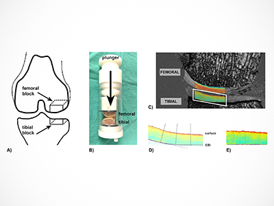
The study, described in a published paper in Osteoarthritis and Cartilage, detailed how Wilson and his colleagues’ used a powerful magnetic resonance imaging (MRI) scanner to determine whether it could measure how weight is distributed in the joint.
“We tried to use MRI to pinpoint where in the joint the load of the body is being transmitted,” he explains. “We currently have no good way of measuring this in living people, and other researchers have had to do very fancy footwork to come up with any measures.”
“We wanted to see if MRI could detect how cartilage changes when it is bearing weight and if the scanner could produce images of the loading in the joint. It’s a novel approach--there are only a handful of groups in the world doing this kind of work.”
To accomplish this, the researchers took pieces of human joints from cadavers, put them in the MRI scanner, and loaded them up in a special test rig. The process was very challenging, admits Wilson.

“It was very complicated and a huge amount of work,” he says, adding that they were able to complete the specialized scan on four samples.
Results from the study were just as complex. The researchers found that the effect of the loading on cartilage was not consistent across the thickness of cartilage in that while the weightbearing part of the cartilage got darker near the surface, cartilage near the bone did not change. While this makes it harder to use the method to measure load, it explains why findings in earlier work have been inconsistent.
“Although our results weren’t as clear or straightforward as we’d hoped, the study does help explain much of what other researchers have seen when they’ve looked at loading up the joints,” says Wilson. “We know now how the images change with depth in the cartilage.”
Building on this research, Wilson and his team have started on a new MRI measure inspired by UBC researchers who work on nerve imaging that has strong potential as to measure load in cartilage.
1 Statistics Canada - Symptom onset, diagnosis and management of osteoarthritis


